Facet syndrome is characterised by Stiffness and pain in the neck with pain radiating to the back of the neck, shoulder and arm for more than 6 months and not responding to medical treatment and physiotherapy, and the primary source of pain was confirmed to be cervical zygapophyseal joints.
Technique:
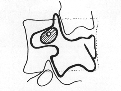
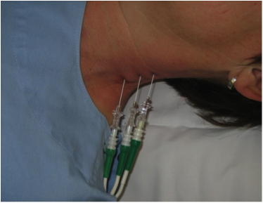
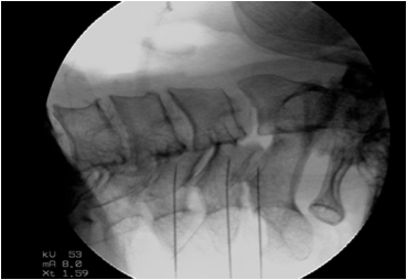
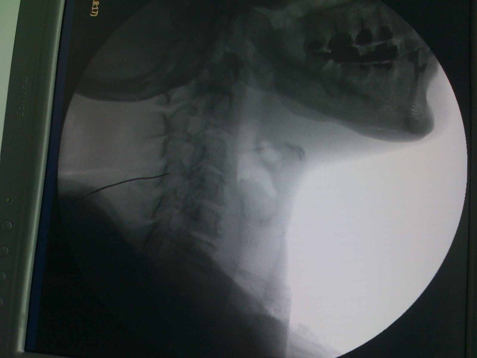
The patient is positioned supine or prone, with the C arm is positioned to visualize the segmental articular pillar and its waist. The insulated RF probe (needle) is directed just medial to the waist of the articular pillar until contacting bone.
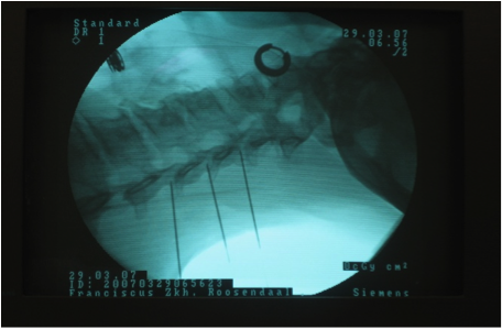
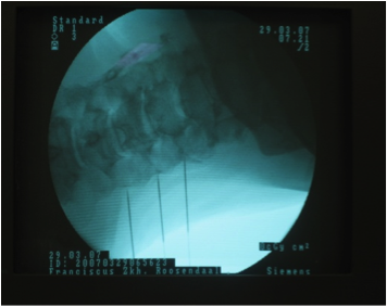
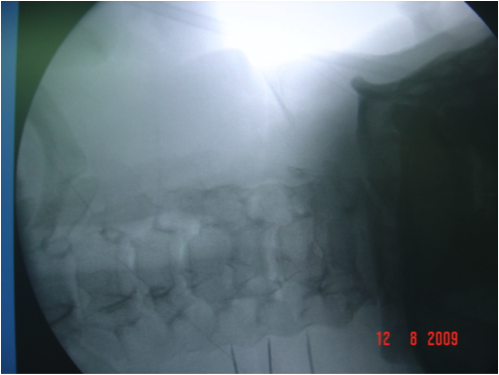
The needle is then slowly redirected just off the articular pillar laterally and repositioned with lateral imaging to the anterior third of the pillar. The Needle placement is confirmed with anterior–posterior (AP) imaging. Sensory and motor stimulation is performed as a safety precaution and to improve the success rate of the procedure.
The voltage at which the patient first perceives the stimulation in the appropriate dermatome is the sensory threshold. This threshold is usually around 0.4–0.7 V when the tip of the needle is next to the medial branch nerve using a frequency of 50 Hz. The frequency is changed to 2 Hz for motor stimulation, and the voltage intensity has to increase to at least twice the sensory threshold before motor activity in the myotomal distribution is typically seen.
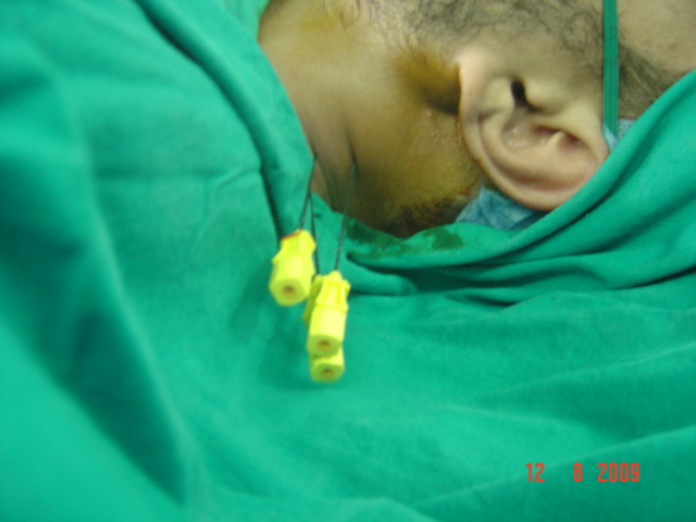
The medial branch nerve is anesthetized before RF lesioning at 80–90°C for 30 s to 2 min per lesion.
The patient is complaining of pain and stiffness in the neck with pain radiating to back of neck, shoulder and arm for more than 6 months. The procedure is performed in an outpatient setting. The treatment is done with local anesthesia along with IV sedation when needed .The patient is positioned supine or prone, with the C arm is positioned to visualize the segmental articular pillar and its waist. The insulated RF probe (needle) is directed just medial to the waist of the articular pillar until contacting bone. The Needle placement is confirmed with anterior–posterior (AP) imaging. Sensory and motor stimulation is performed as a safety precaution and to improve the success rate of the procedure. The medial branch nerve is anesthetized before Radiofrequency lesioning at 80–90°C for 30 s to 2 min per lesion. The needle was removed and puncture site was sterilized and covered.