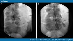
A new minimally invasive diagnostic and therapeutic technique for treatment of chronic spinal pain. Epiduroscopy is a procedure mainly used to allows the visualization of normal anatomical structures, such as the dura mater, blood vessels, connective tissue, nerves and fatty tissue, as well as pathological structures, such as adhesions, inflammatory processes, and fibrosis , although optional interventions such as mechanical or laser mobilization of spinal adhesions, or application of steroids to inflamed tissues, may also be performed.
Technique:
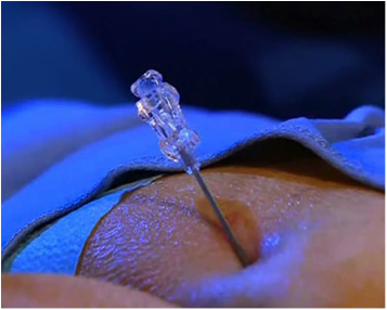
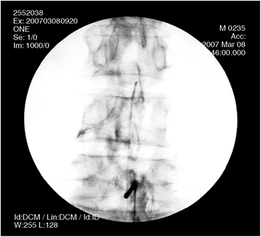
The patient is placed prone, under intravenous sedation and local anesthesia, whilst using X-ray fluoroscopy, Local anesthetic is injected in and around the sacral hiatus or interlaminar to numb the area. A small needle is inserted through the sacral (caudal) hiatus or interlaminar into the epidural space.
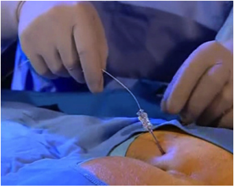
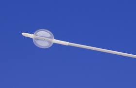
Through this needle is then passed a fine metal guide wire. The small needle is then removed leaving the guide wire in place in the epidural space. A series of dilators are then passed over the guide wire until the sacral membrane will accept a sheath cannula. Once the sheath is in place, the guide wire is removed.
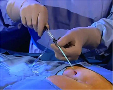
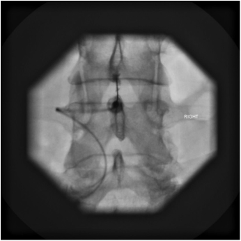
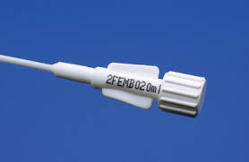
A steerable catheter attached to a fiber optic epiduroscope is then inserted through the center of the sheath until it enters the epidural space. Passage of the steerable catheter is enhanced by using a saline flush system attached to a side port on the sheath. The fiber optic epiduroscope is then advanced upwards using X-ray guidance, until it reaches the area where epidural adhesions have been found on an MRI scan. Epidural adhesions can be gently broken down using the Epiduroscope tip. Afterwards, local anesthetic and steroid can be injected around any inflamed nerve root.